Urethral Stricture Disease

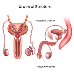
Urethral stricture disorder is a condition characterized by the narrowing or constriction of the urethra, the tube that carries urine from the bladder out of the body. This narrowing can obstruct urine flow and lead to a range of urinary symptoms and complications. Urethral strictures can occur at any age and may result from various causes, manifesting in different types and requiring diverse treatment approaches.

Age Groups:

Urethral stricture disorder can affect individuals of all ages, but it is more commonly diagnosed in men due to their longer urethra. In children, strictures are often congenital or result from trauma. In adults, the condition is frequently related to injury, infection, or previous medical procedures.

Causes:

  • Trauma: Injury to the urethra from accidents or pelvic fractures.
  • Infections: Chronic or severe urinary tract infections, including sexually transmitted infections.
  • Inflammation: Conditions like lichen sclerosus or other inflammatory diseases.
  • Surgical Procedures: Prior surgeries involving the urethra or prostate that result in scar tissue.
  • Congenital Abnormalities: In children, anatomical anomalies present at birth.

Types of Urethral Strictures:

  1. Anterior Urethral Stricture:
    Description
    : Occurs in the part of the urethra located between the external genitalia and the bladder, usually affecting the penile and bulbar urethra.
    Causes: Often related to trauma, infections, or previous surgeries. Common in men and can be caused by procedures like circumcision or injury.
  • Posterior Urethral Stricture:
    Description: Involves the part of the urethra closest to the bladder, including the prostatic and membranous urethra.  Causes: Typically associated with pelvic fractures, prostate surgery, or inflammatory conditions. It is more commonly seen in men and can be linked to chronic conditions like benign prostatic hyperplasia (BPH).
    Symptoms:

  • Difficulty starting urination (hesitancy)
  • Decreased urine stream or flow
  • Painful urination (dysuria)
  • Frequent urination or urgent need to urinate
  • Incomplete bladder emptying
  • Recurrent urinary tract infections
  • Hematuria (blood in the urine)

         Investigations:

  • Uroflowmetry: Measures the rate and pattern of urine flow.
  • Retrograde Urethrogram (RUG): X-ray imaging to visualize the urethra.
  • Cystoscopy: Endoscopic examination to directly view the stricture.
  • Urethral Ultrasound: Provides imaging of the urethra and surrounding tissues.

    Treatments
    :
  • Conservative Management: Includes catheterization or medications to manage symptoms temporarily.
  • Dilation: Gradual widening of the stricture using specialized instruments.
  • Urethrotomy: Surgical procedure to cut and widen the stricture.
  • Urethroplasty: Surgical reconstruction or replacement of the narrowed segment of the urethra.

 Surgery Types:

  • Optical Internal Urethrotomy (OIU): A minimally invasive procedure where a scope is used to incise the stricture.
  • Open Urethroplasty: A more extensive surgery involving the reconstruction of the urethra, often used for complex or recurrent strictures.
  • Buccal Mucosa Graft Urethroplasty: A type of open surgery that uses a graft from the patient’s cheek to repair the stricture.


Conclusion:

Urethral stricture disorder presents with a variety of symptoms and can significantly impact urinary function and quality of life. Accurate diagnosis through imaging and endoscopic evaluations is crucial for determining the appropriate treatment. Management strategies range from minimally invasive procedures to complex reconstructive surgeries, aiming to restore normal urinary function and alleviate symptoms.

Book Appointment

Talk to us?